
What is Liquid Biopsy?
One Visit. Under One hour. From SGD 600.
A plain-language guide to blood-based multi-cancer early detection — what it measures, how the different tests work, and how to read the numbers you will be quoted.
A conventional biopsy takes tissue from a suspected tumour so it can be examined directly. A liquidbiopsy does something far less invasive: it looks for traces of a tumour in a sample of blood. No scan, noradiation, no needle into an organ, no sedation — a single blood draw, analysed in a laboratory.
That idea has moved quickly from research into everyday clinical practice, and MedGlobal Network nowarranges more than one of these tests for clients through partner screening centres in Singapore. Theavailable tests differ from one another in real, technical ways, so this article deliberately does notcompare them. Instead it explains what the whole category does, how the technology works, and whatthe performance figures in the brochures actually mean — so that you can have a better-informedconversation with your doctor.
What a Liquid Biopsy Actually Is
Every cell in the body releases small fragments of its own DNA into the bloodstream as part of an ordinary life cycle. Collectively these are called cell-free DNA, or cfDNA, and everyone has them circulating right now. When a tumour is present, a fraction of that circulating material comes from the tumour itself. That fraction is called circulating tumour DNA, or ctDNA, and it carries the genetic alterations that made the cells cancerous.
A liquid biopsy separates the tumour-derived signal from the ordinary background. Some tests also examine circulating tumour RNA, chemical marks on the DNA, or the physical characteristics of the fragments themselves. When a signal is found, most tests then predict which organ it came from — the tissue-of-origin prediction — so that any follow-up can be targeted precisely rather than becoming a whole-body search.
What Affects How much the Test Can See
The amount of tumour DNA in circulation is not constant. In advanced disease it can be relatively abundant. In the early-stage cancers these tests are designed to find, it is a much smaller fraction of the total circulating DNA. The laboratory is therefore identifying a small number of altered fragments among a great many normal ones — which is precisely the problem the newer sequencing and machine-learning methods were built to solve.
Two practical points follow. Detection generally improves as a tumour grows, so a single quoted sensitivity figure is an average across stages rather than a fixed property of the test. And a result describes what was circulating on the day of the draw, which is why these tests are designed for regular use over time and are intended to work alongside the screening your doctor already recommends, not in place of it.
Not All Liquid Biopsies Read the Same Signal
This is the distinction most often lost in marketing material, and it is the reason two tests can both be liquid biopsies yet produce results that are not directly comparable. Broadly, three kinds of signal are in commercial use, and different tests weight them differently.
Genetic alterations
Reads mutations in tumour DNA at defined targets. Highly precise where a mutation is present, and reports on the specific changes it is designed to detect
Methylation patterns
Reads chemical marks that switch genes on and off. These patterns are strongly tissue-specific, which helps identify where a signal came from.
RNA and fragment features
Reads tumour RNA, or the size andshape of DNA fragments. Living tumour cells release RNA actively, so it can carry signal early.
None of these is straightforwardly superior to the others. They have different strengths across different cancers, and several tests combine more than one. Two useful consequences follow. A larger number of cancers on a panel means broader coverage, not necessarily better detection of any one of them. And sensitivity or specificity figures from different manufacturers are measured on different study populations, so they are best read individually rather than as a league table.
How This Differs from Conventional Tumour Markers
Tests such as PSA, CA-125, CEA and AFP have been in routine use for decades, and they are not liquid biopsies. They measure proteins — substances that rise in the blood when cancer is present, but also in inflammation, benign growths, infection and ordinary individual variation. That is why a raised marker so often leads to investigation that finds nothing wrong.
A liquid biopsy measures tumour genetic material rather than a protein by-product. That is a more specific thing to look for, and it is a large part of why these tests can be designed to raise far fewer unnecessary alarms. It is a genuine difference in kind rather than a marketing distinction. It does not retire the older tests, which remain valuable for monitoring known disease, but for screening purposes it is a meaningful step forward.
Understanding the Numbers: Sensitivity, Specificity, Predictive Value and Accuracy
Every screening test is described by a handful of percentages, and they are easy to mix up because theysound similar. They answer genuinely different questions, and knowing which is which makes any brochure much easier to read.
Sensitivity— among people who do have cancer, the proportion the test correctly flags. Think of itas the catch rate. Higher sensitivity means fewer cancers go unnoticed.
Specificity— among people who do not have cancer, the proportion the test correctly leaves alone.Higher specificity means fewer people are sent for follow-up they did not need.
Positive predictive value— among people who receive a positive result, the proportion who genuinely have cancer. This is the figure that answers the question most people actually ask: what does my result mean for me?
Negative predictive value— among people who receive a negative result, the proportion who genuinely do not have cancer. In screening this is usually the highest of the four figures, and it is the one that applies to most people who take the test.
Accuracy— the proportion of all results, positive and negative together, that were correct. It is the figure most often quoted in advertising and the least informative on its own, for the reason set out below.
A worked illustration
Suppose 10,000 people are screened, and suppose 1 in 100 of them has an undetected cancer. Supposethe test has 80 per cent sensitivity and 99 per cent specificity. Those figures are broadly typical of tests in this category, though every test differs and the numbers below are for illustration only.
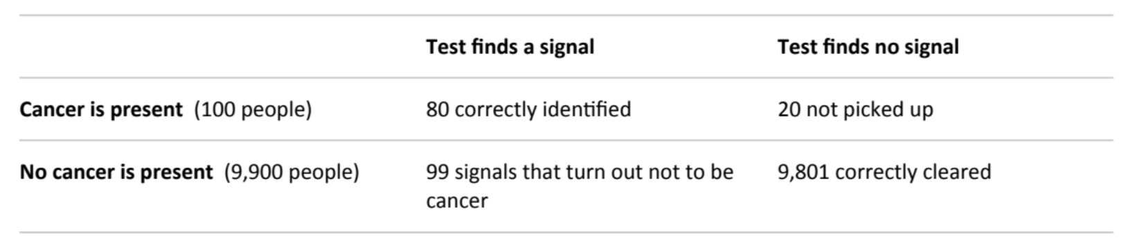
From that single table, all four figures follow:
Sensitivity is 80 per cent — 80 of the 100 cancers were flagged.
Specificity is 99 per cent — 9,801 of the 9,900 people without cancer were correctly cleared.
Positive predictive value is about 45 per cent — of the 179 people who received a signal, 80 had cancer. Roughly one positive result in two turns out to be cancer, which is considerably more informative than several long-established screening tests, where the great majority of positive results are eventually cleared.
Negative predictive value is about 99.8 per cent — of the 9,821 people who received a negative result, 9,801 genuinely had no cancer. This is the outcome the overwhelming majority of people who take the test will receive, and it is strong reassurance.
Accuracy is about 98.8 per cent — 9,881 of the 10,000 results were correct.
That last figure is worth a moment's thought, because it explains why accuracy alone should never be the basis for choosing a test. Almost everyone screened does not have cancer, so a test could correctly clear all of them, miss every single cancer, and still report an accuracy above 98 per cent. Accuracy is dominated by the very large number of correct negatives. Sensitivity, specificity and the two predictive values tell you what is actually happening; accuracy summarises it in a way that flatters every test equally. One further point about predictive value: it depends on how common cancer is in the group being tested, not only on the test itself. The same test produces a higher positive predictive value in a higher-risk group than in a low-risk one. This is why these tests are offered to people with an elevated baseline risk, and why doing so makes each result more meaningful.
What a Result Means in Practice
A negative result is the common outcome and, as the numbers above show, a genuinely reassuring one. It is best understood as a clear reading at a point in time, which is why regular screening is more valuable than a single test, and why it sits alongside the mammograms, colonoscopies and other checks your doctor recommends rather than replacing them.
A positive result is not a diagnosis. It means a cancer signal has been detected and that it warrants a closer look — usually targeted imaging guided by the predicted tissue of origin, which is exactly the information that makes follow-up efficient rather than exhaustive. As with every screening test in medicine, some of those investigations will happily conclude that there is nothing wrong. The value of the test is that when something is found, it tends to be found earlier, when there are more options available.
The practical implication is simply that a good screening arrangement includes a defined next step.Knowing in advance who will interpret the report and what follow-up looks like is what turns a result into a plan.
Who It Suits — and When to Wait
These tests are generally offered to people with an elevated baseline risk rather than to the whole adult population. Typical criteria across providers include being aged 40 and above, having a family history of cancer, or carrying lifestyle or occupational risk factors such as a substantial smoking history.
There are also circumstances in which it is better to wait a little, because other sources of circulating DNA can interfere with a clean reading:
pregnancy;
a cancer diagnosis within the past few years, where the appropriate test is one designed for monitoring rather than screening;
recent surgery, a blood transfusion or a bone marrow transplant.
Exact eligibility and exclusion criteria vary between tests, so these are worth confirming for which ever one you choose. If any of the above applies, postponing the draw for a short period is usually all that is needed.
Further Reading
Multicancer early detection tests at a crossroads: commercial availability ahead of definitive evidence. ASCO Educational Book, 2025. A clinical overview of the category and the questions researchers are still working on.
Clinical implementation of multi-cancer early detection tests: can we find a path forward? Nature Reviews Clinical Oncology,2026.
Wan, J. C. M., Sasieni, P. & Rosenfeld, N. Promises and pitfalls of multi-cancer early detection using liquid biopsy tests.Nature Reviews Clinical Oncology, 2025; 22: 566–580.
Ministry of Health Singapore national screening recommendations. Current recommended ages and intervals can be confirmed via HealthHub or your doctor.
This article is general information and does not constitute medical advice. Multi-cancer early detection testing is intended tocomplement, not replace, the cancer screening recommended for you by a qualified clinician. Figures used in the workedillustration are for explanatory purposes and are not the performance data of any specific test. Product names are the trademarksof their respective owners; no comparison of or endorsement between specific tests is intended.